Description
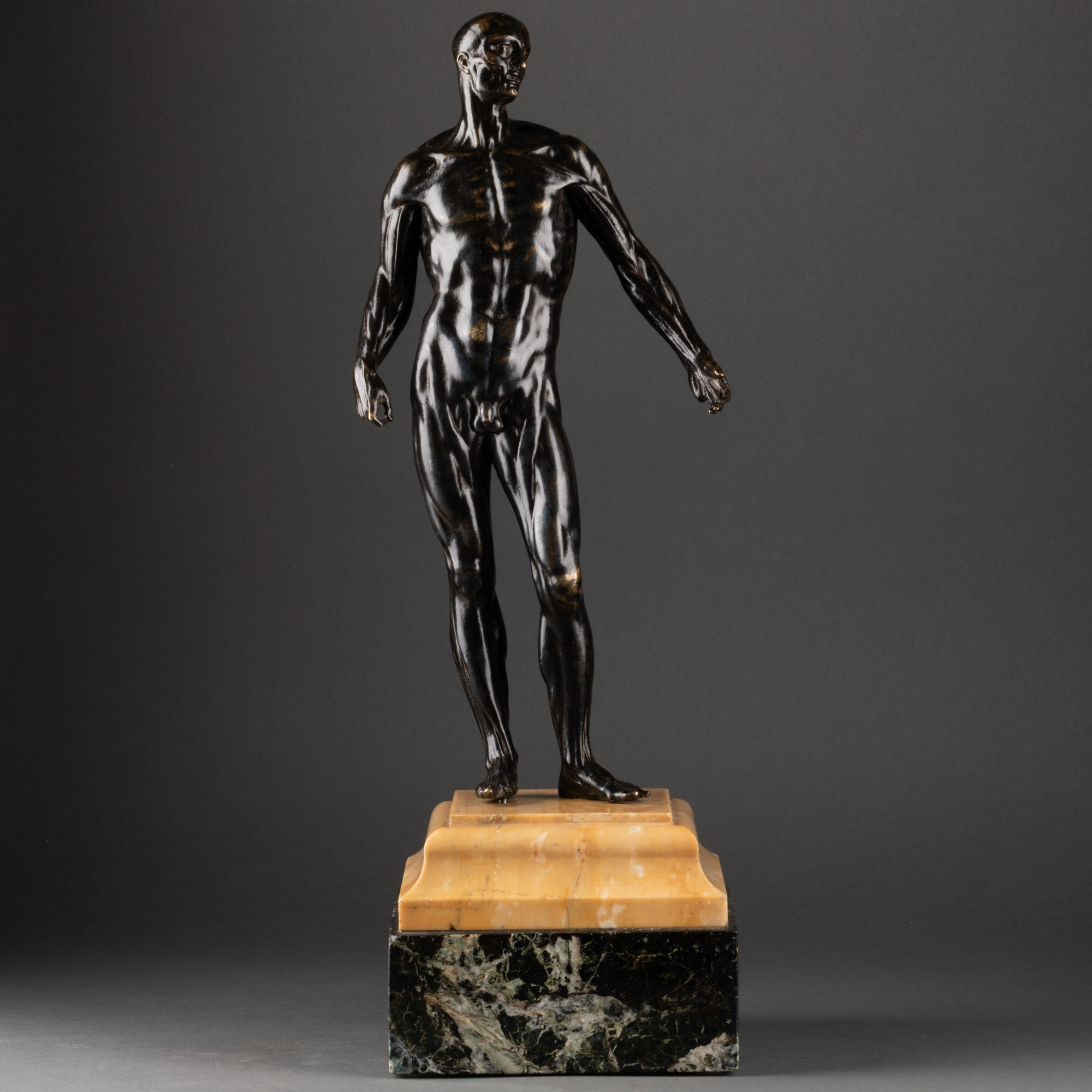
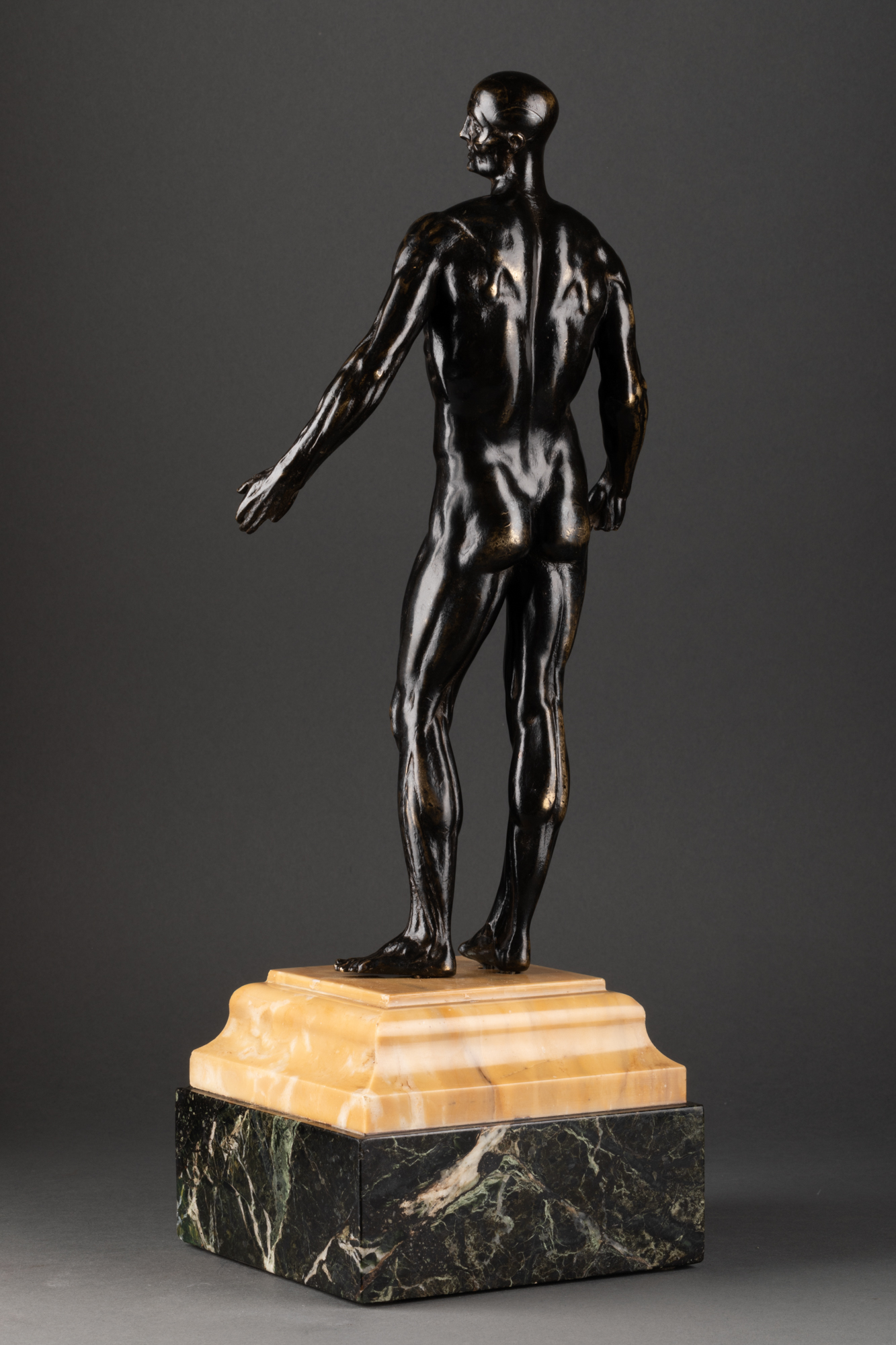
Debout dans un léger contrapposto, l’homme tourne la tête vers sa gauche, son bras droit suit le long du corps avec le pouce et l’index joints, le bras gauche est légèrement ouvert. Ce superbe écorché en bronze repose sur un socle postérieur fait de deux types de marbre : marbre de Sienne et du marbre vert antique.
A partir du XVIe siècle se développe en Italie un intérêt sans précédent pour l’anatomie, mêlant de manière inédite science et art. Cette redécouverte de l’anatomie implique de nombreux changements, notamment concernant la vision quasi « cartoonesque » du corps humain, héritage de la médecine antique. Pendant les siècles précédents, les médecins et physiciens pensaient que les maladies résultaient d’un déséquilibre des quatre humeurs constituant le corps : la sang, la lymphe, la bile jaune et la bile noire. Avec cette vision de la médecine, l’anatomie jouait un rôle très limité dû à une générale incompréhension du rôle des organes, du système de la circulation sanguine et une méconnaissance de la chimie de base. Mais grâce à ce nouvel intérêt pour l’anatomie dès le XIVe siècle et surtout pendant le XVIe, une nouvelle science de la guérison se développe en se basant sur un principe que l’on retrouve dans notre médecine moderne : si un organe est malade alors le traitement doit être appliqué au dit organe plutôt qu’au corps en son intégralité.
Cette révolution scientifique s’observe également, comme bien souvent, au sein de la création artistique. Rappelons qu’à cette époque les manuscrits autrefois recopiés par les religieux se transforment en imprimés et la révolution technologique de l’imprimerie permet une production et diffusion des ouvrages rapide et internationale. Etant une science qui nécessite la description de formes visuelles : l’anatomie a besoin d’images, surtout d’images pouvant être reproduites avec la précision d’une impression. C’est une des raisons qui explique l’implication d’artistes. Si les imprimeurs et les graveurs ont d’abord été les acteurs majeurs de cette étrange et unique collaboration interdisciplinaire ; certains artistes finissent par devenir anatomistes eux-mêmes : leur permettant de développer une réalité anatomique dans leurs créations. Créant, indépendamment de toute médecine, des images anatomiques parfois plus avancées et précises que celles produites par les anatomistes professionnels. Ce n’est qu’au milieu du XVIe siècle que la science « dure » reprend le dessus et obtient le total contrôle sur les illustrations anatomiques.
C’est avec des grands noms comme Michel-Ange et surtout de Vinci que l’intérêt des artistes pour l’anatomie atteint son zénith : artiste et scientifique complet, Léonard réalise de nombreuses découvertes grâces aux dissections, qui lui permettent de réfuter des théories antiques jusque-là en place. Quant à Michel-Ange il étudie l’anatomie exclusivement pour parfaire son art mais pousse sa connaissances du système musculaire à l’extrême. L’étude des muscles et des squelettes devient, à partir de ce moment, une part importante de l’apprentissage des artistes : une bonne connaissance du corps humain pour une meilleure représentation dans l’art.
Comme dit précédemment les scientifiques finissent par assumer un rôle dominant dans l’illustration anatomique. Pour ne citer que lui : Andreas Vasalius, scientifique ayant étudié la médecine à Paris et en Italie, publie en 1543 à Basel, le De humani corporis fabrica(A propos de la fabrique du corps humain). Cet ouvrage est fondateur pour l‘anatomie moderne et participe à l’évolution de la représentation de l’homme et du vivant. Outre son contenu scientifique qui révolutionne la médecine, ce qui rend ce traité exceptionnel est le fait que son auteur, bien que scientifique de formation, supervise les illustrations réalisés pour son ouvrage : alliant ainsi qualité artistique à la rigueur et la précision scientifique. Illustré par plusieurs artistes comme des élèves de Titien, Jan Calcar ou bien Vasalius lui-même, cet ouvrage est encore aujourd’hui considéré comme un des plus beaux livres du monde notamment grâce à la qualité de ses planches d’illustrations. L’une d’entre elle représente un écorché : mis en scène dans un paysage bucolique, un homme debout sans peau, qui laisse donc apparaître tout son système musculaire. Cet ouvrage fondateur marque le début d’une période nouvelle en matière médicale, mais apparait également comme l’aboutissement de cette pensée scientifique datant du XIVe siècle.
La popularité de l’ouvrage se mesure également au nombre de plagiats et copies partielles qui se suivent. Jusqu’à la fin du XVIIe siècle, la majorité des traités d’anatomie comportent des parties de textes et des illustrations copiées ou inspirées de la Fabrica. La planche de l’écorché n’y échappe pas et pour n’en citer qu’un : Gaspar Becerra réalise vers 1556 un écorché très similaire pour illustrer l’ouvrage de l’espagnol Juan de Valverde de Amusco, Historia de la composicion del cuerpo humano. A travers la création et diffusion de ces copies, la représentation de l’écorché se standardise : un homme debout, la tête levée et tournée, un bras ouvert et l’autre tenant le morceau de peau de la cuisse qu’il vient de s’arracher.
D’abord cantonné à la gravure et aux dessins d’illustrations, le thème de l’écorché atteint d’autre domaine de la création artistique avec le temps, notamment les petites statues en bronze qui sont utilisés comme des modèles d’études anatomique pour les élèves et apprentis artistes. Au XVIIIe et XIXe siècle, l’intérêt pour l’écorché évolue : il devient un objet de curiosité. La précision anatomique du système musculaire est parfois diminuée au profit de l’esthétisme, le bronze est privilégié pour créer des objets de qualité et de prestige et surtout les postures peuvent variées (archer relâchant une flèche, écorché philosophant sur sa propre existence, athlète au repos…).
Notre écorché est à placer dans cette période de transition dans la vision, représentation et compréhension du corps humain. Parfait exemple de l’objet qui intriguait les collectionneurs, il est saisissant pour la grande finesse de ciselage et son réalisme.
_____________________________________________
Standing in a slight contrapposto, the man turns his head to his left, his right arm follows along the body with the thumb and index finger joined, the left arm is slightly open. This superb flayed bronze rests on a posterior plinth made of two types of marble: Siena marble and antique green marble. From the 16th century, an unprecedented interest in anatomy developed in Italy, combining science and art in an unprecedented way. This rediscovery of anatomy involves many changes, particularly concerning the almost « cartoonesque » vision of the human body, a legacy of ancient medicine. For centuries, physicians and physicists believed that disease resulted from an imbalance of the four humours that make up the body: blood, lymph, yellow bile and black bile. With this vision of medicine, anatomy played a very limited role due to a general misunderstanding of the role of the organs, the blood circulation system and a misunderstanding of basic chemistry. But thanks to this new interest in anatomy from the 14th century and especially during the 16th century, a new science of healing developed based on a principle found in our modern medicine: if an organ is sick then the treatment must be applied to said organ rather than to the body as a whole. This scientific revolution is also observed, as so often, within artistic creation. It should be remembered that at that time the manuscripts formerly copied by the monks were transformed into printed matter and the technological revolution of the printing press allowed rapid and international production and distribution of works. Being a science that requires the description of visual forms: anatomy needs images, especially images that can be reproduced with the precision of an impression. This is one of the reasons for the involvement of artists. If printers and engravers were initially the major players in this strange and unique interdisciplinary collaboration; some artists end up becoming anatomists themselves: allowing them to develop anatomical reality in their creations. Creating, independent of any medicine, anatomical images that are sometimes more advanced and precise than those produced by professional anatomists. It was not until the middle of the 16th century that “hard” science regained the upper hand and obtained total control over anatomical illustrations. It is with great names like Michelangelo and especially Da Vinci that the interest of artists for anatomy reached its zenith: artist and complete scientist, Leonardo made many discoveries thanks to dissections, which allowed him to refute theories ancient hitherto in place. As for Michelangelo, he studied anatomy exclusively to perfect his art but pushed his knowledge of the muscular system to the extreme. The study of muscles and skeletons becomes, from this moment, an important part of the apprenticeship of artists: a good knowledge of the human body for a better representation in art. As said before scientists end up assuming a dominant role in anatomical illustration. To cite only him: Andreas Vasalius, a scientist who studied medicine in Paris and Italy, published in 1543 in Basel, De humani corporis fabrica (About the making of the human body). This work is a foundation for modern anatomy and participates in the evolution of the representation of man and living things. In addition to its scientific content which revolutionizes medicine, what makes this treatise exceptional is the fact that its author, although a scientist by training, supervises the illustrations produced for his work: thus combining artistic quality with scientific rigor and precision. Illustrated by several artists such as pupils of Titian, Jan Calcar or even Vasalius himself, this work is still considered today as one of the most beautiful books in the world, in particular thanks to the quality of its illustration plates. One of them represents a flayed person: staged in a bucolic landscape, a man standing without skin, which therefore reveals his entire muscular system. This founding work marks the beginning of a new period in medical matters, but also appears as the culmination of this scientific thought dating from the 14th century. The popularity of the book is also measured by the number of plagiarisms and partial copies that follow. Until the end of the 17th century, the majority of anatomical treatises included parts of texts and illustrations copied or inspired by the Fabrica. The board of the Ecorché is no exception and to cite just one: Gaspar Becerra produced a very similar Ecorché around 1556 to illustrate the work of the Spaniard Juan de Valverde de Amusco, Historia de la composicion del cuerpo humano. Through the creation and dissemination of these copies, the representation of the flayed man became standardized: a man standing, his head raised and turned, one arm open and the other holding the piece of skin from the thigh he had just taken off. ‘to tear out. Initially confined to engraving and illustrative drawings, the theme of the skinned person reached other areas of artistic creation over time, notably the small bronze statues which were used as models for anatomical studies for students and apprentice artists. In the 18th and 19th centuries, interest in the écorché evolved: it became an object of curiosity. The anatomical precision of the muscular system is sometimes reduced in favor of aesthetics, bronze is favored to create quality and prestigious objects and above all the postures can vary (archer releasing an arrow, flayed man philosophizing on his own existence, athlete rest…). Our cutaway is to be placed in this period of transition in the vision, representation and understanding of the human body. Perfect example of the object that intrigued collectors, it is striking for the great finesse of the carving and its realism.


